


 The uterine prolapse is a rare postpartum pathology in cows, characterized by the eversion of the uterus through the birth canal, which exposes the endometrium to the external environment1.
 Although predisposing factors for uterine prolapse have been proposed2, its pathophysiological mechanisms remain largely unknown3.
This condition represents a challenge in cattle production, both due to its economic impact and the need for rapid intervention to prevent serious complications such as shock or septicemia1.
|
 The limited availability of recent studies generates variability in therapeutic approaches.
 In this context, the present review details the epidemiology of uterine prolapse in cattle, its predisposing factors and the most recent therapeutic recommendations,
 EPIDEMIOLOGY OF UTERINE PROLAPSES IN CATTLE
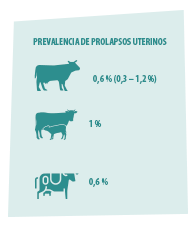 The prevalence of uterine prolapses in cattle shows significant variability, with values ranging between 0.3% and 1.2%2,4-9.
 In this regard, average prevalences of 0.6% have been documented in cattle, with higher frequency of occurrence in beef cows (1%) than in dairy cows (0.6%)7,8.
However, there are other authors who have observed that the highest incidences of uterine prolapses occur in dairy cows10-12.
These discrepancies highlight the need for additional studies to determine the factors of this pathology.
Predisposing factors for uterine prolapses
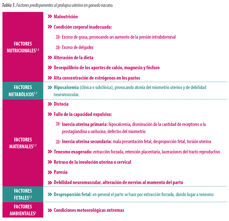
The origin of uterine prolapse is known to be multifactorial (Table 1)1,12:
 It is estimated that 45% of cases are related to dystocia problems, with a higher prevalence in calving of male calves (60.7%).
Hypocalcemia has also been identified as a predisposing factor13,14.
 However, it has not been determined with certainty whether uterine atony is a consequence of hypocalcemia or if, on the contrary, uterine prolapse causes hypocalcemia5,6,15,16.
Unlike vaginal prolapses, no hereditary factors have been identified1,3,12.
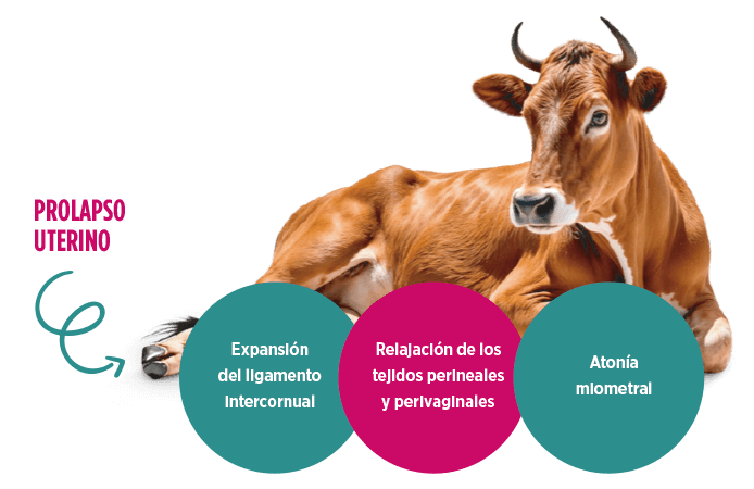
Recipe for a uterine prolapse
It has been reported that uterine prolapse results from the combination of myometrial atony, the expansion of the intercornual ligament and the relaxation of the perineal and perivaginal tissues2.
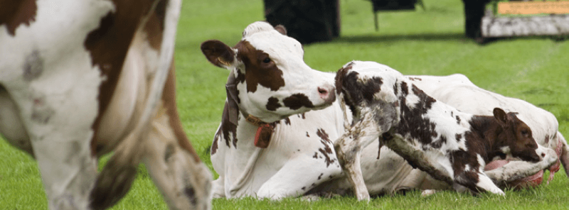
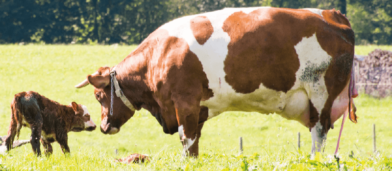
 Generally, it occurs in the first 24 hours postpartum3,12, although cases of prolapse of the non-pregnant horn at the end of gestation have been described1,17.
DIAGNOSIS OF UTERINE PROLAPSE
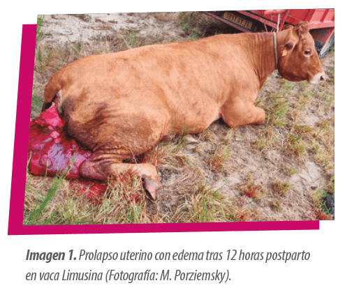
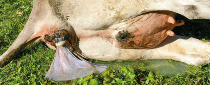
The diagnosis of uterine prolapse is not difficult, as it is characterized by the presence of a large reddish mass with prominent caruncles at the vulva (Image 1).
However, it is essential to evaluate possible concomitant diseases to establish an adequate prognosis and choose the most appropriate treatment.
The therapeutic options include12,18:
 Manual resolution
Hysterectomy
Euthanasia in the most severe cases
1. Preparation prior to the reduction of uterine prolapse
Before proceeding with the resolution of the prolapse, it is recommended to apply:
 Light sedation (xylazine, 0.04-0.06 mg/kg IM or 0.02-0.03 mg/kg IV)20.
Low epidural anesthesia: the dose of anesthetic should not be very high to minimize the risk of falling (1 mL/100 kg body weight of 2% lidocaine)1,3.
In this way, uterine contractions will be reduced, facilitating its manipulation and introduction into the pelvic cavity12. |

2. Washing, reduction of edema and repair of uterine lesions
WASHING
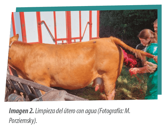
Once the cow is properly anesthetized and immobilized, the greatest amount of fetal membranes should be removed and the endometrium washed with water or saline solution (Image 2). Additionally, placing the uterus in a bag with cold water facilitates its cleaning and reduces the edema.
 It is advisable to apply obstetric lubricant to facilitate its reintroduction3.
EDEMA REDUCTION
For edema reduction, a compressive bandage from distal to proximal can be applied for 10 to 15 minutes.
 Although hypertonic materials, such as sugar, salts, or a 50% dextrose solution effectively reduce uterine edema3,12, they can damage the endometrium22. Therefore, if any of these materials are used, it is recommended to clean and disinfect the uterus after their application1.
Another alternative is to perform a manual massage during repositioning, using an ointment with lubricating and emollient properties23.
INJURY REPAIR
If there is any injury at the uterine level, it can be resolved by simple continuous sutures with catgut #312 or with Reverdin sutures in extensive areas of devitalized tissue12.
Dorsal lacerations 2–3 cm deep do not require suturing, as uterine involution and oxytocin therapy are usually sufficient for resolution21.
3. Resolution of uterine prolapse
REDUCTION OF EDEMA
The repositioning of the uterus is performed by applying pressure on the cervix and uterine body. It is recommended to maintain constant pressure with both closed fists to guide the uterus towards the vagina.
 The use of oven gloves or mittens prevents uterine injuries1,3,12.
 In friable uteri, it is suggested to place them in a plastic bag or damp cloth under compression to reduce the risk of perforation, removing the bandage sequentially during the reduction of the prolapse12.
 A probang, a wiffle ball bat, or a 1.5L bottle can be used to complete the inversion of the gravid horn.
 It is recommended to use warm water to facilitate the complete inversion of the uterine horns, removing the excess by siphoning12.
It is essential to ensure the proper deployment of the uterine horns to prevent reversion or ischemic necrosis1,12.
Cow Position
A Cow standing
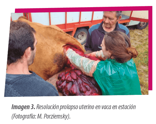
The resolution is more effective when the cow is standing (Image 3), as this12:
Facilitates the alignment of the uterus with the birth canal.
Reduces the risk of contamination
|
B Cow in recumbency
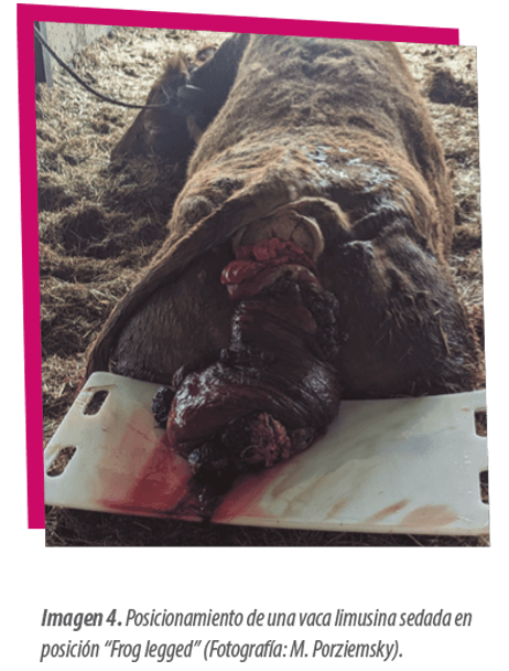
If the cow is in lateral recumbency or according to the operator’s preference, other positions can be applied, such as the “Frog legged” technique (Image 4).
4. Post-treatment after resolution of uterine prolapse
After resolution, administration of oxytocin intramuscularly is indicated to enhance uterine involution, applying:
A Two doses of 20-30 IU with a 30-minute interval3
B A single dose of 20-40 IU12
Although the systematic administration of antibiotics via parenteral route12 was previously recommended, nowadays it is only recommended if the uterus is severely damaged and/or contaminated3.
The placement of retention sutures in the vulva is controversial12:
 If the uterus responds to oxytocin and the cow stands up, prolapse recurrence is unlikely, so suturing is not required3.
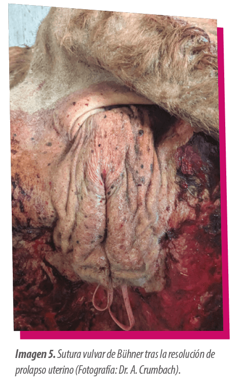
On the other hand, if the cow remains immobile and tenesmus persists, it is recommended to place a vulvar suture for 24-48 hours, with the most common being the Bühner suture (Image 5)1.
PROGNOSIS OF UTERINE PROLAPSE
The prognosis of uterine prolapse is favorable1, depending on early intervention and the absence of concomitant metabolic or musculoskeletal diseases12.
 It is advised to reevaluate the cow within 12-24 hours after resolution1.
The survival rate ranges between 70% and 80%4,8,10,24,25, being higher in nurse cows8.
|
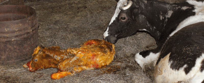
 However, despite the fact that post-resolution conception rates can be over 80%8,24, an increase in the calving-to-calving interval of between 10 and 50 days has been described1,24.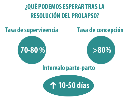
Among the most common complications that can occur after a uterine prolapse, the following stand out1:
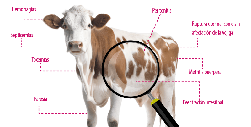
If the cow exhibits tenesmus after an apparently normal resolution, it is recommended to perform an ultrasound of the pelvic canal to detect possible retroflexion of the bladder after uterine repositioning12. |
CONCLUSION
In conclusion, although the average prevalence of uterine prolapses in cows is approximately less than 1%, this pathology has a considerable clinical relevance, with significant economic and animal welfare repercussions.
Despite this reproductive problem being mostly associated with dystocia and hypocalcemia, there are still gaps in the detailed knowledge about its pathophysiological mechanisms, which hinders the implementation of effective preventive plans.
Además, las estrategias terapéuticas actuales presentan áreas de incertidumbre, evidenciadas en maniobras controvertidas como el uso de antibióticos de forma masiva o la aplicación de suturas de retención.
 It is imperative to promote future research that optimizes clinical management and mitigates the impact of this obstetric emergency in the cattle sector.
Acknowledgments: Project PID2023-152404OB-I00 and contract (RYC2021-032245-I), funded by MCIU/ AEI10.13039/501100011033 and FSE+.
BIBLIOGRAPHY
1. Simões, J., Stilwell, G. (2021). Puerperal Complications in the Dam. In: Calving Management and Newborn Calf Care, Cham: Springer. 209–237 https://doi.org/10.1007/978-3-030-68168-5_9
2. Plenderleith, B. (1986). Prolapse of the uterus in the cow. In Practice, 8(1), 14 15. https://doi.org/10.1136/inpract.8.1.14
3. Peter, A. T., King E. H. (2021). Chapter 47: Management of vaginal, cervico vaginal, and uterine prolapse. En: R. M. Hopper, Bovine Reproduction, 2nd ed., Hoboken, NJ: Wiley Blackwell. 571-578.
4. Odegaard, S. (1977). Uterine prolapse in dairy cows. A clinical study with special reference to incidence, recovery and subsequent fertility. Acta Vet. Scand. Suppl., 63, 1–124
5. Patterson, D. J., Bellows, R. A., Burfening, P. J. (1981). Effects of caesarean section, retained placenta, and vaginal or uterine prolapse on subsequent fertility in beef cattle. Journal of Animal Science, 53(4), 916–921. https://doi.org/10.2527/jas1981.534916x
6. Markusfeld, O. (1987). Periparturient traits in seven high dairy herds, incidence rates, association with parity, and interrelationships among traits. Journal of Dairy Science. 70, 158–166. https://doi.org/10.3168/jds.S0022-0302(87)79990-1
7. Correa, M. T., Erb, H. N., Scarlett, J. M. (1992). A nested case-control study of uterine prolapse. Theriogenology, 37(4), 939–945. https://doi.org/10.1016/0093-691x(92)90055-v
8. Carluccio, A., De Amicis, I., Probo, M., Giangaspero, B. A., Veronesi, M. C (2020). Prevalence, survival and subsequent fertility of dairy and beef cows with uterine prolapse. Acta veterinaria Hungarica, 68(1), 91–94. https://doi.org/10.1556/004.2020.00017
9. Martin, A. D., Groseth, P. K., Munthe-Kaas, M., Nødtvedt, A. (2023). Treatment and survival of Norwegian cattle after uterine prolapse. Acta veterinaria Scandinavica, 65(1), 38. https://doi.org/10.1186/s13028-023-00701-1
10. Gardner, I. A., Reynolds, J. P., Risco, C. A., Hird, D. W. (1990). Patterns of uterine prolapse in dairy cows and prognosis after treatment. Journal of the American Veterinary Medical Association, 197(8), 1021–1024.
11. Roine, K., Saloniemi, H. (1978). Incidence of some diseases in connection with parturition in dairy cows. Acta Vet. Scand. 19, 341–353.
12. Miesner, M. D., Anderson, D. E. (2008). Management of uterine and vaginal prolapse in the bovine. The Veterinary clinics of North America. Food animal practice, 24(2), 409–ix. https://doi.org/10.1016/j.cvfa.2008.02.008
13. DeGaris, P. J., Lean I. J. (2008). Milk fever in dairy cows: a review of pathophysiology and control principles, Veterinary journal (London, England: 1997), 176(1), 58–69. https://doi.org/10.1016/j.tvjl.2007.12.029
14. Valldecabres, A., Pires, J. A. A., Silva-Del-Río, N. (2019). Cow-level factors associated with subclinical hypocalcemia at calving in multiparous Jersey cows. Journal of dairy science, 102(9), 8367–8375. https://doi.org/10.3168/jds.2018-16180
15. Richardson, G. F., Klemmer, A. D., Knudsen, D. B. (1981). Observations on uterine prolapse in beef cattle. The Canadian veterinary journal = La revue vétérinaire canadienne, 22(6), 189–191.
16. Risco, C. A., Reynolds, J. P., Hird, D. (1984). Uterine prolapse and hypocalcemia in dairy cows. Journal of the American Veterinary Medical Association, 185(12), 1517–1519.
17. Abdullahi, U. S., Kumi-Diaka, J. (1986). Prolapse of the nongravid horn in a cow with a seven-month pregnancy: A case report. Theriogenology, 26(3), 353–356. https://doi.org/10.1016/0093-691x(86)90154-8
18. Skúladóttir, G., Phythian, C. J., Holmøy, I. H., Myhrene, G., Alvåsen, K., Martin, A. D. (2022). Overview of the practices of on-farm emergency slaughter of cattle in the Nordic countries. Acta veterinaria Scandinavica, 64(1), 9. https://doi.org/10.1186/s13028-022-00627-0
19. Wilson P. J. (1972). A pneumatic appliance for the replacement of the prolapsed uterus of the cow. The Veterinary record, 90(26), 729–730. https://doi.org/10.1136/vr.90.26.729
20. Mullon, P.Y. (2019). General anesthesia, sedation and restraint in cattle. AABP Proceedings, 52(1), 4-6. https://bovine-ojs-tamu.tdl.org/AABP/article/download/7925/7510
21. Hopper, R. M. (2007). Surgical correction of abnormalities of genital organs of cows: Management of uterine prolapse. En: R. S. Youngquist & W. R. Threlfall (Eds.), Current therapy in large animal theriogenology, 2nd ed., Philadelphia: Saunders, Elsevier, 470-471. http://dx.doi.org/10.1016/B978-072169323-1.50063-5
22. Roberts, S. J. (1986). Injuries and diseases of the puperal period. In: Veterinary obstetrics and genital diseases (theriogenology), 3rd ed., Woodstock, VT: Roberts SJ Publisher. 364. https://doi.org/10.1016/0093-691X(86)90160-3
23. Youngquist, R. S. (1989). Bovine uterine prolapse. Theriogenology handbook, Society for Theriogenology, B(3), 79–81.
24. Jubb, T. F., Malmo, J., Brightling, P., Davis, G. M. (1990). Survival and fertility after uterine prolapse in dairy cows. Australian veterinary journal, 67(1), 22–24. https://doi.org/10.1111/j.1751-0813.1990.tb07386.x
25. Murphy, A. M., Dobson, H. (2002). Predisposition, subsequent fertility, and mortality of cows with uterine prolapse. Veterinary Record, 151(24), 733–735.
Continue reading this content
Register for free or log in to access all rumiNews content.