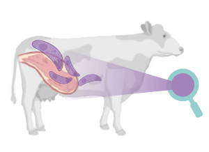
 La neosporosis bovina y la toxoplasmosis ovina son dos enfermedades parasitarias que causan importantes pérdidas económicas en el sector ganadero debido al fallo reproductivo que ocasionan.
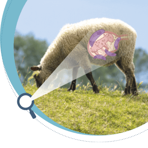 Estas enfermedades están causadas por Neospora caninum y Toxoplasma gondii, respectivamente, ambos son parásitos apicomplejos formadores de quistes estrechamente relacionados, y que comparten muchas características morfológicas y biológicas24,25.
 La neosporosis bovina tiene una gran importancia en el ganado vacuno, ya que es una de las principales causas de aborto a nivel mundial, originando graves pérdidas económicas. El aborto es el principal signo clínico de la neosporosis bovina.
 Los fetos de las madres infectadas por N. caninum pueden morir en el útero y ser reabsorbidos, momificados, sufrir autolisis o nacer muertos.
Las vacas de cualquier edad pueden abortar desde los 3 meses de gestación hasta su término, observándose la mayoría de los abortos a los 5-7 meses de gestación21.
Si la infección ocurre después de la mitad de la gestación pueden nacer terneros vivos con signos clínicos o clínicamente normales, pero persistentemente infectados.
 El fallo reproductivo puede producirse también en otros rumiantes como ovejas, cabras y ciervos21,26,27.
|
 En la actualidad, no existen vacunas ni tratamientos eficaces contra la infección por N. caninum28,29 y las opciones de control se basan en el diagnóstico y en medidas de bioseguridad y manejo21.
Por otro lado, la toxoplasmosis se considera una de las principales causas de pérdidas reproductivas en ovejas y cabras en todo el mundo22,30.
La infección durante la gestación suele provocar la muerte del feto (infección al principio de la gestación); el nacimiento de corderos muertos o débiles, a veces acompañados de un feto de tamaño más pequeño y momificado (infección a mitad de gestación); o el nacimiento de corderos sanos, pero infectados congénitamente (infecciones durante el último tercio de la gestación)31.
Es responsable del 10-23% de los abortos ovinos en Europa y EE.UU.32 y podría ser responsable de entre 680.000 y 1.360.000 abortos anuales en la UE.
La toxoplasmosis es además una importante zoonosis y los pequeños rumiantes pueden desempeñar un papel importante en su transmisión a los humanos33.
 El control de la toxoplasmosis ovina se basa principalmente en la prevención de su transmisión horizontal y en el establecimiento de un programa de vacunación con una cepa viva atenuada S48 (Ovilis Toxovax®).
¿QUÉ TIENEN EN COMÚN N. caninum y T. gondii?
N. caninum y T. gondii comparten muchas características en sus ciclos biológicos, sin embargo, ambos difieren en su rango de hospedadores, el potencial zoonósico (demostrado sólo para T. gondii) y la relevancia de las vías de transmisión horizontal y vertical en el mantenimiento de las infecciones en la naturaleza21,34.
En ambos casos, los hospedadores definitivos son los únicos capaces de producir ooquistes, que son liberados con las heces, mientras que los hospedadores intermediarios desarrollan quistes tisulares repletos de bradizoítos en el tejido muscular y nervioso, donde se cronifica la infección.
|
N. caninum no se considera zoonósico, ya que no se ha detectado la presencia del parásito en los tejidos humanos35.
 La infección postnatal por ingestión de ooquistes (transmisión horizontal) y el paso transplacentario del parásito de la madre al feto (transmisión vertical) son las únicas vías de transmisión demostradas para el ganado y otros hospedadores intermediarios.
A su vez, se han descrito dos formas distintas de transmisión transplacentaria en la neosporosis bovina:
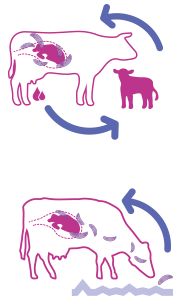 Transmisión transplacentaria endógena: es la más frecuente, y se debe a la reactivación durante la gestación de los bradizoítos presentes en los en las madres persistentemente infectadas.
Transmisión transplacentaria exógena: es menos frecuente, y sucede cuando las madres se infectan por primera vez durante la gestación por ingestión de ooquistes.
La ingestión de bradizoítos contenidos en los quistes tisulares presentes en el hospedador intermediario es la fuente más probable de infección para el hospedador definitivo21.
Por otro lado, los gatos domésticos y otros félidos son los hospedadores definitivos de T. gondii.
La vía más importante de transmisión de T. gondii a los pequeños rumiantes es la horizontal, tras la ingestión oral de ooquistes esporulados que contaminan el forraje o el agua36.
Se estima que sólo el 2% de las ovejas se infectan de forma congénita y que el 4% de las ovejas infectadas de forma persistente transmiten la infección a sus crías37.
También se ha descrito la transmisión transplacentaria endógena del parásito en cabras38.
La interacción de estos parásitos con la placenta es de gran importancia y determina el curso de la infección, pudiendo ocasionar la muerte del feto o la transmisión del parásito a la descendencia. Sin embargo, no se conoce el mecanismo que produce el aborto y todavía hay importantes incógnitas por esclarecer.
|
A continuación, resumiremos las características anatómicas y funcionales de la placenta de los rumiantes en este primer artículo para, en una segunda parte, describir las alternativas de los modelos posibles, junto con sus ventajas e inconvenientes, para el estudio de estos parásitos en la placenta.
LA PLACENTA DE LOS RUMIANTES 
La placenta desempeña un papel fundamental en la patogenia de estas enfermedades, ya que es una barrera biológica que debe atravesar el parásito para infectar al feto. Además, desde el punto de vista inmunológico, en la placenta se produce una respuesta antiinflamatoria que permite:
- La implantación del embrión y garantizar su supervivencia (tolerancia inmunológica).
- A la vez, podría favorecer la multiplicación del parásito en la placenta y su transmisión al feto, comprometiendo su supervivencia.
Al mismo tiempo, las células de la placenta son capaces de defenderse frente a una infección produciendo una respuesta inflamatoria que podría ser potencialmente dañina para el feto e inducir el aborto1.
 A pesar de ello, N. caninum y T. gondii son capaces de replicarse en los tejidos de la placenta, comprometiendo el éxito de la gestación.
En los mamíferos, el crecimiento y la supervivencia del feto dependen de la placenta, un órgano temporal formado por un entramado de tejidos maternos y fetales que mantiene a ambos individuos conectados (Figura 1).
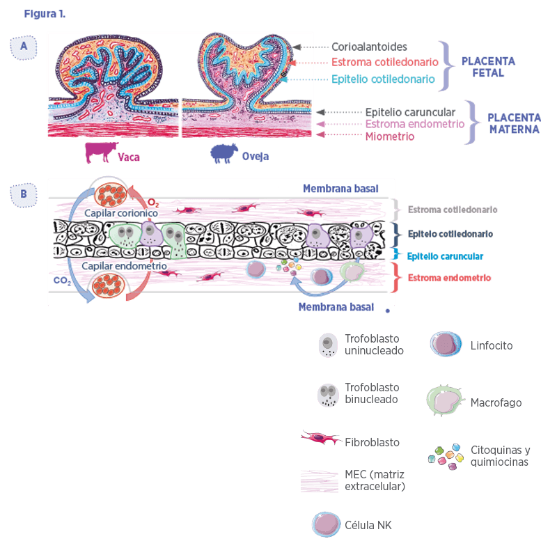

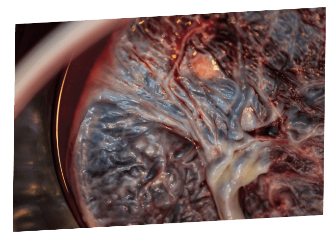
 Los rumiantes domésticos presentan placentas corioalantoideas, vellosas y cotiledonarias.
Estos términos se refieren a la fusión del corion y el alantoides en zonas discretas de la placenta fetal para formar proyecciones vellosas denominadas cotiledones.
En los rumiantes, el proceso de implantación comienza a los 16 (oveja) o 20 (vaca) días tras la cubrición, cuando el epitelio coriónico del embrión (formado por células del trofoblasto) desarrolla los cotiledones de origen fetal, los cuales se entrelazan con las carúnculas, cuyo origen es el endometrio materno.
La combinación de un cotiledón fetal y una carúncula materna se denomina placentoma, la unidad funcional de la placenta de los rumiantes (Figura 1A)3,6.
Por otra parte, las células del trofoblasto son componentes clave de la placenta, describiéndose dos poblaciones diferentes:
 Células uninucleadas: forman la mayor parte de la interfaz feto-maternal y participan principalmente en el intercambio de nutrientes
 Células binucleadas: participan en la síntesis de hormonas como el lactógeno placentario y la progesterona7,8 (Figura 1B)
La matriz extracelular (MEC) es otro componente importante de la placenta:
- Participa en la adhesión del trofoblasto al endometrio uterino Induce la diferenciación celular9
- Actúa como reservorio de factores de crecimiento10
- Sirve como vía por la que las células pueden migrar11
La MEC consiste en una mezcla de proteoglicanos, glicosaminoglicanos y componentes estructurales como diferentes tipos de colágeno, fibronectina y laminina, que son sintetizados principalmente por los fibroblastos.
Estas células también producen metaloproteinasas de la matriz (MMP), que degradan los componentes de la MEC y están reguladas por inhibidores tisulares de MMP; el desequilibrio de estas proteínas se ha asociado a diferentes condiciones patológicas y a la diseminación de determinados patógenos, entre ellos T. gondii, y de células infectadas a través de la barrera placentaria12,13.
|
En cuanto a sus funciones, la placenta:
Forma una interfaz entre la circulación materna y la fetal, actuando así como sistema digestivo, respiratorio, de excreción, metabólico y endocrino del feto.
Secreta las hormonas necesarias para el mantenimiento de la gestación, la adaptación del metabolismo materno, el crecimiento fetal, el parto e incluso la lactancia.
Constituye una barrera física que protege al feto contra los gérmenes y fármacos que puedan circular por el torrente sanguíneo materno.
Sin embargo, algunos patógenos son capaces de atravesar esta barrera e infectar al feto debido a su reducido tamaño (p.ej., los virus), o a su capacidad de replicarse tanto en las células placentarias maternas como en las fetales (por ejemplo, T. gondii, N. caninum, Brucella abortus o Chlamydia abortus).
|

 No obstante, la modulación inmunitaria local que tiene lugar durante la gestación es un proceso complejo que todavía no se conoce completamente.
Leer Interacciones placenta-patógeno en parásitos apicomplejos de rumiantes – Parte II
Adaptado y traducido del artículo original: “Modeling the ruminant placenta-pathogen interactions in apicomplexan parasites: current and future perspectives”, Pastor-Fernández I, Collantes-Fernández E, Jiménez-Pelayo L, Ortega-Mora LM and Horcajo P (2021); Front. Vet. Sci. 7:634458. doi: 10.3389/ fvets.2020.634458
1. Rindsjö E, Holmlund U, Sverremark-Ekström E, Papadogiannakis N, Scheynius A. Toll-like receptor-2 expression in normal and pathologic human placenta. Hum Pathol. (2007) 38:468–73. doi: 10.1016/j.humpath.2006.09.009
2. Innes EA, Bartley PM, Maley SW, Wright SE, Buxton D. Comparative host parasite relationships in ovine toxoplasmosis and bovine neosporosis and strategies for vaccination. Vaccine. (2007) 25:5495–503. doi:10.1016/j.vaccine.2007.02.044
3. Peter AT. Bovine placenta: a review on morphology, components, and defects from terminology and clinical perspectives. Theriogenology. (2013) 80:693–705. doi: 10.1016/j.theriogenology.2013.06.004
4. Haeger J-, Hambruch N, Pfarrer C. The bovine placenta in vivo and in vitro. Theriogenology. (2016) 86:306–12. doi: 10.1016/j.theriogenology.2016.04.043
5. Fowden AL, Ward JW, Wooding FB, Forhead AJ. Developmental programming of the ovine placenta. Soc Reprod Fertil Suppl. (2010) 67:41–57 doi: 10.5661/RDR-VII-41
6. Wooding F. The synepitheliochorial placenta of ruminants: binucleate cell fusions and hormone production. Placenta. (1992) 13:101–13. doi: 10.1016/0143-4004(92)90025-O
7. Duello TM, Byatt JC, Bremel RD. Immunohistochemical localization of placental lactogen in binucleate cells of bovine placentomes. Endocrinology. (1986) 119:1351–5. doi: 10.1210/endo-119-3-1351
8. Reimers TJ, Ullmann MB, Hansel W. Progesterone and prostanoid production by bovine binucleate trophoblastic cells. Biol Reprod. (1985) 33:1227–36. doi: 10.1095/biolreprod33.5.1227
9. Nakano H, Shimada A, Imai K, Takezawa T, Takahashi T, Hashizume K. Bovine trophoblastic cell differentiation on collagen substrata: formation of binucleate cells expressing placental lactogen. Cell Tissue Res. (2002) 307:225–35. doi: 10.1007/s00441-001-0491-x
10. Taipale J, Keski-Oja J. Growth factors in the extracellular matrix. FASEB J. (1997) 11:51–9. doi: 10.1096/fasebj.11.1.9034166
11. Pfarrer C, Hirsch P, Guillomot M, Leiser R. Interaction of integrin receptors with extracellular matrix is involved in trophoblast giant cell migration in bovine placentomes. Placenta. (2003) 24:588–97. doi: 10.1016/S0143-4004(03)00059-6
12. Wang M, Lai S. Fibronectin degradation by MMP-2/MMP-9 in the serum of pregnant women and umbilical cord with Toxoplasma gondii infection. J Obstet Gynaecol. (2013) 33:370–4. doi: 10.3109/01443615.2013.769501
13. Brasil TR, Freire-de-Lima CG, Morrot A, Vetö Arnholdt AC. Host-Toxoplasma gondii coadaptation leads to fine tuning of the immune response. Front Immunol. (2017) 8:1080. doi: 10.3389/fimmu.2017.01080
14. Donnelly L, Campling G. Functions of the placenta. Anaesth Intens Care. (2016) 17:349–53. doi: 10.1016/j.mpaic.2016.04.004
15. Schuler G, Greven H, Kowalewski MP, Doring B, Ozalp GR, Hoffmann B. Placental steroids in cattle: hormones, placental growth factors or by-products of trophoblast giant cell differentiation? Exp Clin Endocrinol Diabetes. (2008) 116:429–36. doi: 10.1055/s-2008-1042408
16. Jiménez-Pelayo L, García-Sánchez M, Collantes-Fernández E, Regidor-Cerrillo J, Horcajo P, GutiérrezExpósito D, et al. The cross-talk at the placenta between Neospora caninum and the bovine host determines the outcome of the infection. Vet Res. (2020) 51:83. doi: 10.1186/s13567-020-00803-y
17. Castaño P, Fernandez M, Regidor-Cerrillo J, Fuertes M, Horcajo P, Ferre I, et al. Peripheral and placental immune responses in sheep after experimental infection with Toxoplasma gondii at the three terms of gestation. Vet Res. (2019) 50:66. doi: 10.1186/s13567-019-0681-8
18. Arranz-Solís D, Benavides J, Regidor-Cerrillo J, Horcajo P, Castaño P, del Carmen Ferreras M, et al. Systemic and local immune responses in sheep after Neospora caninum experimental infection at early, mid and late gestation. Vet Res. (2016) 47:1–13. doi: 10.1186/s13567-015-0290-0
19. Wegmann TG, Lin H, Guilbert L, Mosmann TR. Bidirectional cytokine interactions in the maternalfetal relationship: is successful pregnancy a TH2 phenomenon? Immunol Today. (1993) 14:353–6. doi: 10.1016/0167-5699(93)90235-D
20. Entrican G. Immune regulation during pregnancy and host-pathogen interactions in infectious abortion. J Comp Pathol. (2002) 126:79–94. doi: 10.1053/jcpa.2001.0539
21. Dubey JP, Hemphill A, Calero-Bernal R, Schares G. Neosporosis in Animals. Boca Raton, FL: CRC Press (2017). doi: 10.1201/9781315152561
22. Stelzer S, Basso W, Silván JB, Ortega-Mora L, Maksimov P, Gethmann J, et al. Toxoplasma gondii infection and toxoplasmosis in farm animals: Risk factors and economic impact. Food Waterborne Parasitol. (2019):e00037. doi: 10.1016/j.fawpar.2019.e00037
23. Pappas G, Roussos N, Falagas ME. Toxoplasmosis snapshots: global status of Toxoplasma gondii seroprevalence and implications for pregnancy and congenital toxoplasmosis. Int J Parasitol. (2009) 39:1385–94. doi: 10.1016/j.ijpara.2009.04.003
24. Speer CA, Dubey JP, McAllister MM, Blixt JA. Comparative ultrastructure of tachyzoites, bradyzoites, and tissue cysts of Neospora caninum and Toxoplasma gondii. Int J Parasitol. (1999) 29:1509–19. doi: 10.1016/S0020-7519(99)00132-0
25. Reid AJ, Vermont SJ, Cotton JA, Harris D, Hill-Cawthorne GA, Konen Waisman S, et al. Comparative genomics of the apicomplexan parasites Toxoplasma gondii and Neospora caninum: Coccidia differing in host range and transmission strategy. PLoS Pathog. (2012) 8:e1002567. doi: 10.1371/journal.ppat.1002567
26. González-Warleta M, Castro-Hermida JA, Calvo C, Pérez V, Gutiérrez-Expósito D, Regidor-Cerrillo J, et al. Endogenous transplacental transmission of Neospora caninum during successive pregnancies across three generations of naturally infected sheep. Vet Res. (2018) 49:106. doi: 10.1186/s13567-018-0601-3
27. Costa RC, Mesquita LP, Nunes MVL, Oliveira IM, Oliveira LFS, Souza AR, et al. Neospora caninum bioassay in gerbils using placental tissues from naturally infected goats. Vet Parasitol. (2018) 249:70–3. doi: 10.1016/j.vetpar.2017.11.009
28. Horcajo P, Regidor-Cerrillo J, Aguado-Martínez A, Hemphill A, Ortega-Mora LM. Vaccines for bovine neosporosis: current status and key aspects for development. Parasite Immunol. (2016) 38:709–23. doi: 10.1111/pim.12342
29. Sánchez-Sánchez R, Vázquez P, Ferre I, Ortega-Mora LM. Treatment of toxoplasmosis and neosporosis in farm ruminants: state of knowledge and future trends. Curr Top Med Chem. (2018). doi: 10.2174/1568026618666181002113617
30. Dubey JP, Murata FHA, Cerqueira-Cezar CK, Kwok OCH. Public health and economic importance of Toxoplasma gondii infections in goats: the last decade. Res Vet Sci. (2020) 132:292–307. doi: 10.1016/j. rvsc.2020.06.014
31. Trees AJ, Williams DJL. Endogenous and exogenous transplacental infection in Neospora caninum and Toxoplasma gondii. Trends Parasitol. (2005) 21:558–61. doi: 10.1016/j.pt.2005.09.005
32. Dubey JP, Su C. Population biology of Toxoplasma gondii: what’s out and where did they come from. Mem Inst Oswaldo Cruz. (2009) 104:190–5. doi: 10.1590/S0074-02762009000200011
33. Belluco S, Mancin M, Conficoni D, Simonato G, Pietrobelli M, Ricci A. Investigating the determinants of Toxoplasma gondii prevalence in meat: a systematic review and meta-regression. PLoS ONE. (2016) 11:153856. doi: 10.1371/journal.pone.0153856
34. Dubey JP. Toxoplasmosis of Animals and Humans. Boca Raton, FL: CRC Press (2016). doi: 10.1201/9781420092370
35. Calero-Bernal R, Horcajo P, Hernández M, Ortega-Mora LM, Fuentes I. Absence of Neospora caninum DNA in Human Clinical Samples, Spain. Emerg Infect Dis. (2019) 25:1226–7. doi: 10.3201/eid2506.181431
36. Elmore SA, Jones JL, Conrad PA, Patton S, Lindsay DS, Dubey JP. Toxoplasma gondii: epidemiology, feline clinical aspects, and prevention. Trends Parasitol. (2010) 26:190–6. doi: 10.1016/j.pt.2010.01.009
37. Innes EA, Bartley PM, Buxton D, Katzer F. Ovine toxoplasmosis. Parasitology. (2009) 136:1887–94. doi: 10.1017/S0031182009991636
38. Dubey JP. Repeat transplacental transfer of Toxoplasma gondii in dairy goats. J Am Vet Med Assoc. (1982) 180:1220–1.
39. Dos Santos TR, Faria Gabriela da SM, Guerreiro BM, Lopes WDZ, da Silva HM, Garcia JL, et al. Congenital toxoplasmosis in chronically infected and subsequently challenged ewes. PLoS ONE. (2016) 11:e0165124. doi: 10.1371/journal.pone.0165124
40. Klauck V, Pazinato R, Radavelli WM, Custodio E, Bianchi AE, Camillo G, et al. Toxoplasma gondii infection in dairy ewes: vertical transmission and influence on milk production and reproductive performance. Microb Pathog. (2016) 99:101–5. doi: 10.1016/j.micpath.2016.08.012
41. Dubey JP, Lindsay DS. Neosporosis, toxoplasmosis, and sarcocystosis in ruminants. Vet Clin North Am Food Anim Pract. (2006) 22:645–71. doi: 10.1016/j.cvfa.2006.08.001
42. Innes EA. The host-parasite relationship in pregnant cattle infected with Neospora caninum. Parasitology. (2007) 134:1903–10. doi: 10.1017/S0031182007000194
43. Quinn HE, Ellis JT, Smith NC. Neospora caninum: a cause of immune mediated failure of pregnancy? Trends Parasitol. (2002) 18:391–4. doi: 10.1016/S1471-4922(02)02324-3
44. Buxton D, Finlayson J. Experimental infection of pregnant sheep with Toxoplasma gondii: pathological and immunological observations on the placenta and fetus. J Comp Pathol. (1986) 96:319–33. doi: 10.1016/0021-9975(86)90052-6
45. Collantes-Fernández E, Arnaiz-Seco I, Burgos BM, Rodríguez-Bertos A, Aduriz G, Fernández-García A, et al. Comparison of Neospora caninum distribution, parasite loads and lesions between epidemic and endemic bovine abortion cases. Vet Parasitol. (2006) 142:187–91. doi: 10.1016/j.vetpar.2006.05.030
46. Benavides J, Collantes-Fernández E, Ferre I, Perez V, Campero C, Mota R, et al. Experimental ruminant models for bovine neosporosis: what is known and what is needed. Parasitology. (2014) 141:1471–88. doi: 10.1017/S0031182014000638
47. Castaño P, Fuertes M, Regidor-Cerrillo J, Ferre I, Fernández M, Ferreras MC, et al. Experimental ovine toxoplasmosis: influence of the gestational stage on the clinical course, lesion development and parasite distribution. Vet Res. (2016) 47:1. doi: 10.1186/s13567-016-0327-z
48. Regidor-Cerrillo J, Álvarez-García G, Pastor-Fernández I, Marugán Hernández V, Gómez-Bautista M, Ortega-Mora LM. Proteome expression changes among virulent and attenuated Neospora caninum isolates. J Proteomics. (2012) 75:2306–18 doi: 10.1016/j.jprot.2012.01.039
49. Regidor-Cerrillo J, Gómez-Bautista M, Sodupe I, Aduriz G, Álvarez-García G, Del Pozo I, et al. In vitro invasion efficiency and intracellular proliferation rate comprise virulence-related phenotypic traits of Neospora caninum. Vet Res. (2011) 42:41. doi: 10.1186/1297-9716-42-41
50. Dellarupe A, Regidor-Cerrillo J, Jiménez-Ruiz E, Schares G, Unzaga JM, Venturini MC, et al. Comparison of host cell invasion and proliferation among Neospora caninum isolates obtained from oocysts and from clinical cases of naturally infected dogs. Exp Parasitol. (2014) 145:22–8. doi: 10.1016/j. exppara.2014.07.003
51. Dellarupe A, Regidor-Cerrillo J, Jiménez-Ruiz E, Schares G, Unzaga JM, Venturini MC, et al. Clinical outcome and vertical transmission variability among canine Neospora caninum isolates in a pregnant mouse model of infection. Parasitology. (2014) 141:356–66. doi: 10.1017/S0031182013001479
52. Rojo-Montejo S, Collantes-Fernández E, Blanco-Murcia J, Rodríguez-Bertos A, Risco-Castillo V, Ortega-Mora LM. Experimental infection with a low virulence isolate of Neospora caninum at 70 days gestation in cattle did not result in foetopathy. Vet Res. (2009) 40:49. doi: 10.1051/vetres/2009032
53. Jiménez-Pelayo L, García-Sánchez M, Vázquez P, Regidor-Cerrillo J, Horcajo P, Collantes-Fernández E, et al. Early Neospora caninum infection dynamics in cattle after inoculation at mid-gestation with high (Nc-Spain7)- or low (Nc Spain1H)-virulence isolates. Vet Res. (2019) 50:72,019-0691-6. doi: 10.1186/ s13567-019-0691-6.
54. Sibley LD, Boothroyd JC. Virulent strains of Toxoplasma gondii comprise a single clonal lineage. Nature. (1992) 359:82. doi: 10.1038/359082a0
55. Saeij JP, Boyle JP, Boothroyd JC. Differences among the three major strains of Toxoplasma gondii and their specific interactions with the infected host. Trends Parasitol. (2005) 21:476–81. doi: 10.1016/j. pt.2005.08.001
56. Jiménez-Pelayo L, García-Sánchez M, Regidor-Cerrillo J, Horcajo P, Collantes Fernández E, GómezBautista M, et al. Differential susceptibility of bovine caruncular and trophoblast cell lines to infection with high and low virulence isolates of Neospora caninum. Parasit Vectors. (2017) 10:463. doi: 10.1186/ s13071-017-2409-9
57. Jiménez-Pelayo L, García-Sánchez M, Regidor-Cerrillo J, Horcajo P, Collantes Fernández E, GómezBautista M, et al. Immune response profile of caruncular and trophoblast cell lines infected by high- (NcSpain7) and low virulente (Nc-Spain1H) isolates of Neospora caninum. Parasit Vectors. (2019) 12(1):218. doi: 10.1186/s13071-019-3466-z
58. Horcajo P, Jiménez-Pelayo L, García-Sánchez M, Regidor-Cerrillo J, Collantes Fernández E, Rozas D, et al. Transcriptome modulation of bovine trophoblast cells in vitro by Neospora caninum. Int J Parasitol. (2017) 47:791–9. doi: 10.1016/j.ijpara.2017.08.007
59. Geurts N, Opdenakker G, Van den Steen PE. Matrix metalloproteinases as therapeutic targets in protozoan parasitic infections. Pharmacol Ther. (2012) 133:257–79. doi: 10.1016/j.pharmthera.2011.11.008
60. Huppertz B, Kivity V, Sammar M, Grimpel Y, Leepaz N, Orendi K, et al. Cryogenic and low temperature preservation of human placental villous explants – a new way to explore drugs in pregnancy disorders. Placenta. (2011) 32(Suppl.):S65–76. doi: 10.1016/j.placenta.2010.11.020
61. Quintanilla-Gozalo A, Pereira-Bueno J, Seijas-Carballedo A, Costas E, Ortega Mora LM. Observational studies in Neospora caninum infected dairy cattle: relationship infection-abortion and gestational antibody fluctuations. In: Hemphill A, Gottstein B. A European perspective on Neospora caninum. Int J Parasitol. (2000) 30:877–924. doi: 10.1016/S0020-7519(00)00072-2
62. Guy CS, Williams DJL, Kelly DF, McGarry JW, Guy F, Bjorkman C, et al. Neospora caninum in persistently infected, pregnant cows: spontaneous transplacental infection is associated with an acute increase in maternal antibody. Vet Rec. (2001) 149:443–9. doi: 10.1136/vr.149.15.443
63. Rosbottom A, Gibney H, Kaiser P, Hartley C, Smith RF, Robinson R, et al. Up regulation of the maternal immune response in the placenta of cattle naturally infected with Neospora caninum. PLoS ONE. (2011) 6:e15799. doi: 10.1371/journal.pone.0015799
64. Sánchez-Sánchez R, Ferre I, Regidor-Cerrillo J, Gutiérrez-Expósito D, Ferrer LM, Arteche-Villasol N, et al. Virulence in mice of a Toxoplasma gondii type II isolate does not correlate with the outcome of experimental infection in pregnant sheep. Front Cell Infect Microbiol. (2019) 8:436. doi: 10.3389/ fcimb.2018.00436
65. Yarovinsky F, Sher A. Toll-like receptor recognition of Toxoplasma gondii. Int J Parasitol. (2006) 36:255–9. doi: 10.1016/j.ijpara.2005.12.003
66. Koblansky AA, Jankovic D, Oh H, Hieny S, Sungnak W, Mathur R, et al. Recognition of profilin by Toll-like receptor 12 is critical for host resistance to Toxoplasma gondii. Immunity. (2013) 38:119–30. doi: 10.1016/j.immuni.2012.09.016
67. Ivanova DL, Fatima R, Gigley JP. Comparative analysis of conventional natural killer cell responses to acute infection with Toxoplasma gondii strains of different virulence. Front Immunol. (2016) 7:347. doi: 10.3389/fimmu.2016.00347
68. Wattegedera SR, Doull LE, Goncheva MI, Wheelhouse NM, Watson DM, Pearce J, et al. Immunological homeostasis at the ovine placenta may reflect the degree of maternal fetal interaction. Front Immunol. (2019) 9:3025. doi: 10.3389/fimmu.2018.03025
69. Castaño P, Fuertes M, Fernández M, Ferreras MC, Ferre I, Ortega-Mora LM, et al. Macrophages and T Lymphocytes in the ovine placenta after experimental infection with Toxoplasma gondii. Vet Pathol. (2020) 57:545–9. doi: 10.1177/0300985820923987
70. Quinn HE, Miller CM, Ellis JT. The cell-mediated immune response to Neospora caninum during pregnancy in the mouse is associated with a bias toward production of interleukin-4. Int J Parasitol. (2004) 34:723–32. doi: 10.1016/j.ijpara.2004.01.007
71. Almería S, Serrano-Pérez B, Darwich L, Mur-Novales R, Garcia-Ispierto I, Cabezón O, et al. Cytokine gene expression in aborting and non-aborting dams and in their fetuses after experimental infection with Neospora caninum at 110 days of gestation. Vet Parasitol. (2016) 227:138–42. doi: 10.1016/j. vetpar.2016.08.006
72. Mur-Novales R, Serrano-Pérez B, García-Ispierto I, de Sousa NM, Beckers JF, Almería S, et al. Experimental Neospora caninum infection modifies trophoblast cell populations and plasma pregnancyassociated glycoprotein 1 and 2 dynamics in pregnant dairy heifers. Vet Parasitol. (2016) 216:7–12. doi: 10.1016/j.vetpar.2015.12.001
73. Fernández-Escobar M, Millán J, Chirife AD, Ortega-Mora LM, Calero-Bernal R. Molecular survey for cyst-forming coccidia (Toxoplasma gondii, Neospora caninum, Sarcocystis spp.) in Mediterranean periurban micromammals. Parasitol Res. (2020) 119:2679–86. doi: 10.1007/s00436-020-06777-2
74. Mévélec M, Ducournau C, Ismael AB, Olivier M, Sèche É, Lebrun M, et al. Mic1 3 Knockout Toxoplasma gondii is a good candidate for a vaccine against T. gondii induced abortion in sheep. Vet Res. (2010) 41:49. doi: 10.1051/vetres/2010021
75. McCann CM, McAllister MM, Gondim LF, Smith RF, Cripps PJ, Kipar A, et al. Neospora caninum in cattle: Experimental infection with oocysts can result in exogenous transplacental infection, but not endogenous transplacental infection in the subsequent pregnancy. Int J Parasitol. (2007) 37:1631–9. doi: 10.1016/j.ijpara.2007.05.012
76. Caspe SG, Moore DP, Leunda MR, Cano DB, Lischinsky L, Regidor-Cerrillo J, et al. The Neospora caninum-Spain 7 isolate induces placental damage, fetal death and abortion in cattle when inoculated in early gestation. Vet Parasitol. (2012) 189:171–81. doi: 10.1016/j.vetpar.2012.04.034
77. Regidor-Cerrillo J, Arranz-Solís D, Benavides J, Gómez-Bautista M, Castro Hermida JA, Mezo M, et al. Neospora caninum infection during early pregnancy in cattle: how the isolate influences infection dynamics, clinical outcome and peripheral and local immune responses. Vet Res. (2014) 45:10. doi: 10.1186/1297-9716-45-10
78. Almería S, Serrano-Perez B, Darwich L, Domingo M, Mur-Novales R, Regidor Cerrillo J, et al. Fetal death in naive heifers inoculated with Neospora caninum isolate Nc-Spain7 at 110 days of pregnancy. Exp Parasitol. (2016) 168:62–9. doi: 10.1016/j.exppara.2016.06.009
79. Macaldowie C, Maley SW, Wright S, Bartley P, Esteban-Redondo I, Buxton D, et al. Placental pathology associated with fetal death in cattle inoculated with Neospora caninum by two different routes in early pregnancy. J Comp Pathol. (2004) 131:142–56. doi: 10.1016/j.jcpa.2004.02.005
80. Maley SW, Buxton D, Rae AG, Wright SE, Schock A, Bartley PM, et al. The pathogenesis of neosporosis in pregnant cattle: inoculation at mid-gestation. J Comp Pathol. (2003) 129:186–95. doi: 10.1016/S0021-9975(03)00032-X
81. Vázquez P, Osoro K, Fernández M, Román-Trufero A, Regidor-Cerrillo J, Jiménez-Pelayo L, et al. Effects of challenge dose and inoculation route of the virulent Neospora caninum Nc-Spain7 isolate in pregnant cattle at mid-gestation. Vet Res. (2019) 50:68. doi: 10.1186/s13567-019-0700-9
82. Castaño P, Fuertes M, Ferre I, Fernández M, del Carmen Ferreras M, Moreno Gonzalo J, et al. Placental thrombosis in acute phase abortions during experimental Toxoplasma gondii infection in sheep. Vet Res. (2014) 45:9. doi: 10.1186/1297-9716-45-9
83. Dubey JP, Murata FHA, Cerqueira-Cezar CK, Kwok OCH, Su C. Economic and public health importance of Toxoplasma gondii infections in sheep: 2009–2020. Vet Parasitol. (2020) 286:109195. doi: 10.1016/j.vetpar.2020.109195
84. Mavin S, Joss AW, Ball J, Ho-Yen DO. Do Toxoplasma gondii RH strain tachyzoites evolve during continuous passage? J Clin Pathol. (2004) 57:609–11. doi: 10.1136/jcp.2003.013763
85. Khan A, Behnke MS, Dunay IR, White MW, Sibley LD. Phenotypic and gene expression changes among clonal type I strains of Toxoplasma gondii. Eukaryot Cell. (2009) 8:1828–36. doi: 10.1128/ EC.00150-09
86. Regidor-Cerrillo J, Xia D, Jiménez-Pelayo L, García-Sánchez M, Collantes Fernández E, Randle N, et al. Proteomic characterization of host-pathogen interactions during bovine trophoblast cell line infection by Neospora caninum. Pathogens. (2020) 9:749. doi: 10.3390/pathogens9090749
87. Rosbottom A, Gibney EH, Guy CS, Kipar A, Smith RF, Kaiser P, et al. Upregulation of cytokines is detected in the placentas of cattle infected with Neospora caninum and is more marked early in gestation when fetal death is observed. Infect Immun. (2008) 76:2352–61. doi: 10.1128/IAI.01780-06
88. Gibney EH, Kipar A, Rosbottom A, Guy CS, Smith RF, Hetzel U, et al. The extent of parasiteassociated necrosis in the placenta and fetal tissues of cattle following Neospora caninum infection in early and late gestation correlates with fetal death. Int J Parasitol. (2008) 38:579–88. doi: 10.1016/j.ijpara.2007.09.015
89. Canton GJ, Katzer F, Maley SW, Bartley PM, Benavides-Silvan J, Palarea Albaladejo J, et al. Inflammatory infiltration into placentas of Neospora caninum challenged cattle correlates with clinical outcome of pregnancy. Vet Res. (2014) 45:11. doi: 10.1186/1297-9716-45-11
90. Marin MS, Hecker YP, Quintana S, Pérez S, Leunda MR, Cantón G, et al. Immunization with inactivated antigens of Neospora caninum induces toll-like receptors 3, 7, 8 and 9 in maternal-fetal interface of infected pregnant heifers. Vet Parasitol. (2017) 243:12–7. doi: 10.1016/j.vetpar.2017.06.005
91. Maley SW, Buxton D, Macaldowie CN, Anderson IE, Wright SE, Bartley PM, et al. Characterization of the immune response in the placenta of cattle experimentally infected with Neospora caninum in early gestation. J Comp Pathol. (2006) 135:130–41. doi: 10.1016/j.jcpa.2006.07.001
92. Cantón GJ, Katzer F, Maley SW, Bartley PM, Benavides-Silván J, Palarea Albaladejo J, et al. Cytokine expression in the placenta of pregnant cattle after inoculation with Neospora caninum. Vet Immunol Immunopathol. (2014) 161:77–89. doi: 10.1016/j.vetimm.2014.07.004
93. Almería S, Araujo RN, Darwich L, Dubey JP, Gasbarre LC. Cytokine gene expression at the maternofetal interface after experimental Neospora caninum infection of heifers at 110 days of gestation. Parasite Immunol. (2011) 33:517–23. doi: 10.1111/j.1365-3024.2011.01307.x
94. Serrano-Pérez B, Hansen PJ, Mur-Novales R, Garcia-Ispierto I, de Sousa NM, Beckers JF, et al. Crosstalk between uterine serpin (SERPINA14) and pregnancy associated glycoproteins at the fetal-maternal interface in pregnant dairy heifers experimentally infected with Neospora caninum. Theriogenology. (2016) 86:824–30. doi: 10.1016/j.theriogenology.2016.03.003
95. Almería S, Araujo R, Tuo W, López-Gatius F, Dubey JP, Gasbarre LC. Fetal death in cows experimentally infected with Neospora caninum at 110 days of gestation. Vet Parasitol. (2010) 169(3-4):304–11. doi:10.1016/j.vetpar.2009.12.044
96. Almería S, Serrano-Pérez B, Darwich L, Araujo R, López-Gatius F, Dubey J, et al. Maternal and fetal immune response patterns in heifers experimentally infected with Neospora caninum in the second trimester of pregnancy–a descriptive study. Vet Parasitol. (2014) 204:146–52. doi: 10.1016/j.vetpar.2014.05.006
97. Hecker YP, Cantón G, Regidor-Cerrillo J, Chianini F, Morrell E, Lischinsky L, et al. Cell mediated immune responses in the placenta following challenge of vaccinated pregnant heifers with Neospora caninum. Vet Parasitol. (2015) 214:247–54. doi: 10.1016/j.vetpar.2015.10.015
98. Dorsch M, de Yaniz M, Fiorani F, Hecker Y, Odeón A, Morrell E, et al. A descriptive study of lectin histochemistry of the placenta in cattle following inoculation of Neospora caninum. J Comp Pathol. (2019) 166:45–53. doi: 10.1016/j.jcpa.2018.10.172
99. Horcajo P, Pitarch A, Jiménez-Pelayo L, García-Sánchez M, Collantes Fernández E, Ortega-Mora LM. Comparative proteomic analysis of bovine placentas infected with high and low virulence Neospora caninum isolates. in Proceedings of the 5th International Meeting on Apicowplexan parasites in Farm Animals. Berlin. (2019).
100. Benavides J, Fernández M, Castaño P, Ferreras M, Ortega-Mora L, Pérez V. Ovine toxoplasmosis: a new look at its pathogenesis. J Comp Pathol. (2017) 157:34–8. doi: 10.1016/j.jcpa.2017.04.003
101. Trees AJ, Crozier SJ, Buxton D, Blewett DA. Serodiagnosis of ovine toxoplasmosis: an assessment of the latex agglutination test and the value of IgM specific titres after experimental oocyst-induced infections. Res Vet Sci. (1989) 46:67–72. doi: 10.1016/S0034-5288(18)31120-2
102. Owen MR, Clarkson MJ, Trees AJ. Acute phase Toxoplasma abortions in sheep. Vet Rec. (1998) 142:480–2. doi: 10.1136/vr.142.18.480
103. Gutiérrez-Expósito D, Arteche-Villasol N, Vallejo-Garcia R, Ferreras-Estrada MC, Ferre I, SánchezSánchez R, et al. Characterization of fetal brain damage in early abortions of ovine toxoplasmosis. Vet Pathol. (2020) 57:535–44. doi: 10.1177/0300985820921539
104. Orendi K, Kivity V, Sammar M, Grimpel Y, Gonen R, Meiri H, et al. Placental and trophoblastic in vitro models to study preventive and therapeutic agents for preeclampsia. Placenta. (2011) 32(Suppl.):S49–54. doi: 10.1016/j.placenta.2010.11.023
105. Stringfellow D, Gray B, Lauerman L, Thomson M, Rhodes P, Bird R. Monolayer culture of cells originating from apreimplantation bovine embryo. In vitro Cell Dev Biol. (1987) 23:750–4. doi: 10.1007/BF02623675
106. Talbot NC, Caperna TJ, Edwards JL, Garrett W, Wells KD, Ealy AD. Bovine blastocyst-derived trophectoderm and endoderm cell cultures: interferon tau and transferrin expression as respective in vitro markers. Biol Reprod. (2000) 62:235–47. doi: 10.1095/biolreprod62.2.235
107. Talbot NC, Powell AM, Ocon OM, Caperna TJ, Camp M, Garrett WM, et al. Comparison of the interferon-tau expression from primary trophectoderm outgrowths derived from IVP, NT, and parthenogenote bovine blastocysts. Mol Reprod Dev. (2008) 75:299–308. doi: 10.1002/mrd.20741
108. Shimada A, Nakano H, Takahashi T, Imai K, Hashizume K. Isolation and characterization of a bovine blastocyst-derived trophoblastic cell line, BT-1: development of a culture system in the absence of feeder cell. Placenta. (2001) 22:652–62. doi: 10.1053/plac.2001.0702
109. Ramos-Ibeas P, Calle A, Pericuesta E, Laguna-Barraza R, Moros-Mora R, Lopera-Vásquez R, et al. An efficient system to establish biopsy-derived trophoblastic cell lines from bovine embryos. Biol Reprod. (2014) 91:15. doi: 10.1095/biolreprod.114.118430
110. Suzuki Y, Koshi K, Imai K, Takahashi T, Kizaki K, Hashizume K. Bone morphogenetic protein 4 accelerates the establishment of bovine trophoblastic cell lines. Reproduction. (2011) 142:733–43. doi: 10.1530/REP-11-0275
111. Pillai VV, Siqueira LG, Das M, Kei TG, Tu LN, Herren AW, et al. Physiological profile of undifferentiated bovine blastocyst-derived trophoblasts. Biol Open. (2019) 8:10.1242/bio.037937. doi: 10.1242/bio.037937
112. Saadeldin IM, Abdelfattah-Hassan A, Swelum AA. Feeder cell type affects the growth of in vitro cultured bovine trophoblast cells. Biomed Res Int. (2017) 2017:1061589. doi: 10.1155/2017/1061589
113. Kawaguchi T, Cho D, Hayashi M, Tsukiyama T, Kimura K, Matsuyama S, et al. Derivation of induced trophoblast cell lines in cattle by doxycycline-inducible piggyBac vectors. PLoS ONE. (2016) 11:e0167550. doi:10.1371/journal.pone.0167550
114. Munson L, Chandler SK, Schlafer DH. Long-term culture of bovine trophoblastic cells. J Tissue Cult Methods. (1988) 11:123–8. doi: 10.1007/BF01404264
115. Vanselow J, Fürbass R, Tiemann U. Cultured bovine trophoblast cells differentially express genes encoding key steroid synthesis enzymes. Placenta. (2008) 29:531–8. doi: 10.1016/j.placenta.2008.03.004
116. Hambruch N, Haeger JD, Dilly M, Pfarrer C. EGF stimulates proliferation in the bovine placental trophoblast cell line F3 via Ras and MAPK. Placenta. (2010) 31:67–74. doi: 10.1016/j.placenta.2009.10.011
117. Martino N, Rizzo A, Pizzi F, Dell’Aquila M, Sciorsci R. Effects of kisspeptin-10 on in vitro proliferation and kisspeptin receptor expression in primary epithelial cell cultures isolated from bovine placental cotyledons of fetuses at the first trimester of pregnancy. Theriogenology. (2015) 83:978–987. doi:10.1016/j.theriogenology.2014.11.033
118. Bridger P, Haupt S, Klisch K, Leiser R, Tinneberg H, Pfarrer C. Validation of primary epitheloid cell cultures isolated from bovine placental caruncles and cotyledons. Theriogenology. (2007) 68:592–603. doi:10.1016/j.theriogenology.2007.05.046
119. Bridger PS, Menge C, Leiser R, Tinneberg HR, Pfarrer CD. Bovine caruncular epithelial cell line (BCEC-1) isolated from the placenta forms a functional epithelial barrier in a polarized cell culture model. Placenta. (2007) 28:1110–7. doi: 10.1016/j.placenta.2007.07.002
120. Wango EO, Heap RB, Wooding FB. Progesterone and 5 beta-pregnanediol production by isolated fetal placental binucleate cells from sheep and goats. J Endocrinol. (1991) 129:283–9. doi: 10.1677/joe.0.1290283
121. Morgan G, Whyte A, Wooding FB. Characterization of the synthetic capacities of isolated placental binucleate cells from sheep and goats. Anat Rec. (1990) 226:27–36. doi: 10.1002/ar.1092260105
122. Soares MC, Servely JL, Puissant C, Bolifraud P, Lacroix MC, Schaeffer B, et al. Ovine chorionic somatomammotrophin (oCS) production by isolated cotyledon cells from sheep in early and midgestation: auto-regulation by recombinant oCS. J Endocrinol. (1999) 161:289–98. doi: 10.1677/joe.0.1610289
123. Steven DH, Mallon KA, Nathanielsz PW. Sheep trophoblast in monolayer cell culture. Placenta. (1980) 1:209–21. doi: 10.1016/S0143-4004(80)80003-8
124. Farmer JL, Burghardt RC, Jousan FD, Hansen PJ, Bazer FW, Spencer TE. Galectin 15 (LGALS15) functions in trophectoderm migration and attachment. FASEB J. (2008) 22:548–60. doi: 10.1096/fj.07-9308com
125. Ralph MM, Lee CS, Thorburn GD. Identification and characterization of monolayer cultures of sheep trophoblast cells maintained in bicameral culture chambers. Biol Reprod. (1989) 41:481–9. doi: 10.1095/biolreprod41.3.481
126. Kim J, Song G, Gao H, Farmer JL, Satterfield MC, Burghardt RC, et al. Insulin-like growth factor II activates phosphatidylinositol 3-kinase protooncogenic protein kinase 1 and mitogen-activated protein kinase cell Signaling pathways, and stimulates migration of ovine trophectoderm cells. Endocrinology. (2008) 149:3085–94. doi: 10.1210/en.2007-1367
127. Haldorson GJ, Stanton JB, Mathison BA, Suarez CE, Baszler TV. Neospora caninum: antibodies directed against tachyzoite surface protein NcSRS2 inhibit parasite attachment and invasion of placental trophoblasts in vitro. Exp Parasitol. (2006) 112:172–8. doi: 10.1016/j.exppara.2005.11.004
128. Zhang Y, Shi J, Liu S. Establishment and characterization of a telomerase immortalized sheep trophoblast cell line. Biomed Res Int. (2016) 2016:5808575. doi: 10.1155/2016/5808575
129. Jimenéz-Meléndez A, Fernández-Álvarez M, Calle A, Ramírez MA, Diezma Diaz C, VázquezArbaizar P, et al. Lytic cycle of Besnoitia besnoiti tachyzoites displays similar features in primary bovine endothelial cells and fibroblasts. Parasit Vectors. (2019) 12:517. doi: 10.1186/s13071-019-3777-0
130. Hemphill A, Gottstein B. Neospora caninum and neosporosis – recent achievements in host and parasite cell biology and treatment. Acta Parasitol (2006) 51:15–25. doi: 10.2478/s11686-006-0002-z
131. Almería S, Serrano-Pérez B, López-Gatius F. Immune response in bovine neosporosis: protection or contribution to the pathogenesis of abortion. Microbial Pathogenesis. (2017) 109:177–82. doi: 10.1016/j.micpath.2017.05.042
132. Pinheiro AM, Costa SL, Freire SM, Ribeiro CS, Tardy M, El-Bacha RS, et al. Neospora caninum: early immune response of rat mixed glial cultures after tachyzoites infection. Exp Parasitol. (2010) 124:442–7. doi:10.1016/j.exppara.2009.12.018
133. Xu T, Ping J, Yu Y, Yu F, Yu Y, Hao P, et al. Revealing parasite influence in metabolic pathways in Apicomplexa infected patients. BMC Bioinformatics. (2010) 11(Suppl.11):S13. doi:10.1186/1471-2105-11-S11-S13
134. Remmerie A, Scott CL. Macrophages and lipid metabolism. Cell Immunol (2018) 330:27–42. doi: 10.1016/j.cellimm.2018.01.020
135. Hargrave KE, Woods S, Millington O, Chalmers S, Westrop GD, Roberts CW. Multi-omics studies demonstrate Toxoplasma gondii-induced metabolic reprogramming of murine dendritic cells. Front Cell Infect Microbiol. (2019) 9:309 doi: 10.3389/fcimb.2019.00309
136. Nelson MM, Jones AR, Carmen JC, Sinai AP, Burchmore R, Wastling JM. Modulation of the host cell proteome by the intracellular apicomplexan parasite Toxoplasma gondii. Infect Immun. (2008) 76:828–44. doi: 10.1128/IAI.01115-07
137. Pernas L, Adomako-Ankomah Y, Shastri AJ, Ewald SE, Treeck M, Boyle JP, et al. Toxoplasma effector MAF1 mediates recruitment of host mitochondria and impacts the host response. PLoS Biol. (2014) 12:e1001845. doi: 10.1371/journal.pbio.1001845
138. Nolan SJ, Romano JD, Luechtefeld T, Coppens I. Neospora caninum recruits host cell structures to its parasitophorous vacuole and salvages lipids from organelles. Eukaryot Cell. (2015) 14:454–73. doi: 10.1128/EC.00262-14
139. Syn G, Anderson D, Blackwell JM, Jamieson SE. Toxoplasma gondii infection is associated with mitochondrial dysfunction in-vitro. Front Cell Infect Microbiol. (2017) 7:512. doi: 10.3389/fcimb.2017.00512
140. García-Sánchez M, Jiménez-Pelayo L, Horcajo P, Regidor-Cerrillo J, Ólafsson EB, Bhandage AK, et al. Differential responses of bovine monocyte-derived macrophages to infection by Neospora caninum isolates of high and low virulence. Front Immunol. (2019) 10:915. doi: 10.3389/fimmu.2019.00915
141. Moreno B, Collantes-Fernández E, Villa A, Navarro A, Regidor-Cerrillo J, Ortega-Mora LM. Occurrence of Neospora caninum and Toxoplasma gondii infections in ovine and caprine abortions. Vet Parasitol. (2012) 187:312–8. doi: 10.1016/j.vetpar.2011.12.034
142. Wheelhouse N, Wattegedera S, Stanton J, Maley S, Watson D, Jepson C, et al. Ovine trophoblast is a primary source of TNFα during Chlamydophila abortus infection. J Reprod Immunol. (2009) 80:49–56. doi: 10.1016/j.jri.2008.12.003
143. Wheelhouse N, Coyle C, Barlow PG, Mitchell S, Greub G, Baszler T, et al. Waddlia chondrophila infects and multiplies in ovine trophoblast cells stimulating an inflammatory immune response. PLoS ONE. (2014) 9:e102386. doi:10.1371/journal.pone.0102386
144. Castro AS, Alves CM, Angeloni MB, Gomes AO, Barbosa BF, Franco PS, et al. Trophoblast cells are able to regulate monocyte activity to control Toxoplasma gondii infection. Placenta. (2013) 34:240–7. doi: 10.1016/j.placenta.2012.12.006
145. da Silva RJ, Gomes AO, Franco PS, Pereira AS, Milian ICB, Ribeiro M, et al. Enrofloxacin and toltrazuril are able to reduce Toxoplasma gondii growth in human BeWo trophoblastic cells and villous explants from human third trimester pregnancy. Front Cell Infect Microbiol. (2017) 7:340. doi: 10.3389/ fcimb.2017.00340
146. Angeloni MB, Guirelli PM, Franco PS, Barbosa BF, Gomes AO, Castro AS, et al. Differential apoptosis in BeWo cells after infection with highly (RH) or moderately (ME49) virulent strains of Toxoplasma gondii is related to the cytokine profile secreted, the death receptor Fas expression and phosphorylated ERK1/2 expression. Placenta. (2013) 34:973–82. doi: 10.1016/j.placenta.2013.09.005
147. Huckle W. Cell-and tissue-based models for study of placental development. Prog Mol Biol Transl Sci. (2017) 145:29–37. doi: 10.1016/bs.pmbts.2016.12.002
148. Miller RK, Genbacev O, Turner MA, Aplin JD, Caniggia I, Huppertz B. Human placental explants in culture: approaches and assessments. Placenta. (2005) 26:439–48. doi: 10.1016/j.placenta.2004.10.002
149. Ferro EAV, Mineo JR, Ietta F, Bechi N, Romagnoli R, Silva DAO, et al. Macrophage migration inhibitory factor is up-regulated in human first-trimester placenta stimulated by soluble antigen of Toxoplasma gondii, resulting in increased monocyte adhesion on villous explants. Am J Pathol. (2008) 72:50–8. doi: 10.2353/ajpath.2008.070432
150. de Oliveira Gomes A, de Oliveira Silva, Deise Aparecida, Silva NM, de Freitas Barbosa B, Franco PS, Angeloni MB, et al. Effect of macrophage migration inhibitory factor (MIF) in human placental explants infected with Toxoplasma gondii depends on gestational age. Am J Pathol. (2011) 178:2792–801. doi:10.1016/j.ajpath.2011.02.005
151. Robbins JR, Zeldovich VB, Poukchanski A, Boothroyd JC, Bakardjiev AI. Tissue barriers of the human placenta to infection with Toxoplasma gondii. Infect Immun. (2012) 80:418–28. doi: 10.1128/IAI.05899-11
152. Ander SE, Rudzki EN, Arora N, Sadovsky Y, Coyne CB, Boyle JP. Human placental syncytiotrophoblasts restrict Toxoplasma gondii attachment and replication and respond to infection by producing immunomodulatory chemokines. mBio. (2018) 9:e01678–17. doi: 10.1128/mBio.01678-17
153. Almeida MPO, Ferro EAV, Briceno MPP, Oliveira MC, Barbosa BF, Silva NM. Susceptibility of human villous (BeWo) and extravillous (HTR-8/SVneo) trophoblast cells to Toxoplasma gondii infection is modulated by intracellular iron availability. Parasitol Res. (2019) 118:1559–72. doi: 10.1007/s00436-019-06257-2
154. Castro-Filice LS, Barbosa BF, Angeloni MB, Silva NM, Gomes AO, Alves CM, et al. Azithromycin is able to control Toxoplasma gondii infection in human villous explants. J Transl Med. (2014) 12:132. doi:10.1186/1479-5876-12-132
155. Franco PS, Gois PSG, de Araujo TE, da Silva RJ, de Freitas Barbosa B, de Oliveira Gomes A, et al. Brazilian strains of Toxoplasma gondii are controlled by azithromycin and modulate cytokine production in human placental explants. J Biomed Sci. (2019) 26:10. doi: 10.1186/s12929-019-0503-3
156. Castillo C, Muñoz L, Carrillo I, Liempi A, Gallardo C, Galanti N, et al. Ex vivo infection of human placental chorionic villi explants with Trypanosoma cruzi and Toxoplasma gondii induces different Tolllike receptor expression and cytokine/chemokine profiles. Am J Reprod Immunol. (2017) 78:12660. doi:10.1111/aji.12660
157. Liempi A, Castillo C, Medina L, Rojas M, Maya JD, Parraguez VH, et al. Ex vivo infection of human placental explants with Trypanosoma cruzi and Toxoplasma gondii: differential activation of NF kappa B signaling pathways. Acta Trop. (2019) 199:105153. doi: 10.1016/j.actatropica.2019.105153
158. Luján CD, Triquell MF, Sembaj A, Guerrero CE, Fretes RE. Trypanosoma cruzi: productive infection is not allowed by chorionic villous explant from normal human placenta in vitro. Exp Parasitol. (2004) 108:176–81. doi:10.1016/j.exppara.2004.07.013
159. Triquell MF, Díaz-Luján C, Freilij H, Paglini P, Fretes RE. Placental infection by two subpopulations of Trypanosoma cruzi is conditioned by differential survival of the parasite in a deleterious placental medium and not by tissue reproduction. Trans R Soc Trop Med Hyg. (2009) 103:1011–8. doi: 10.1016/j. trstmh.2009.03.004
160. Díaz-Luján C, Triquell MF, Castillo C, Hardisson D, Kemmerling U, Fretes RE. Role of placental barrier integrity in infection by Trypanosoma cruzi. Acta Trop. (2016) 164:360–8. doi: 10.1016/j.actatropica.2016.09.021
161. Sartori MJ, Pons P, Mezzano L, Lin S, de Fabro SP. Trypanosoma cruzi infection induces microfilament depletion in human placenta syncytiotrophoblast. Placenta. (2003) 24:767–71. doi: 10.1016/S0143- 4004(03)00111-5
162. Duaso J, Rojo G, Cabrera G, Galanti N, Bosco C, Maya JD, et al. Trypanosoma cruzi induces tissue disorganization and destruction of chorionic villi in an ex vivo infection model of human placenta. Placenta. (2010) 31:705–11. doi:10.1016/j.placenta.2010.05.007
163. Duaso J, Rojo G, Jana F, Galanti N, Cabrera G, Bosco C, et al. Trypanosoma cruzi induces apoptosis in ex vivo infected human chorionic villi. Placenta. (2011) 32:356–61. doi: 10.1016/j.placenta.2011.02.005
164. Liempi A, Castillo C, Duaso J, Droguett D, Sandoval A, Barahona K, et al. Trypanosoma cruzi induces trophoblast differentiation: a potential local antiparasitic mechanism of the human placenta? Placenta. (2014) 35:1035–42. doi: 10.1016/j.placenta.2014.09.017
165. Castillo C, Muñoz L, Carrillo I, Liempi A, Medina L, Galanti N, et al. Toll-like receptor-2 mediates local innate immune response against Trypanosoma cruzi in ex vivo infected human placental chorionic villi explants. Placenta. (2017) 60:40–6. doi: 10.1016/j.placenta.2017.10.005
166. Fievet N, Moussa M, Tami G, Maubert B, Cot M, Deloron P, et al. Plasmodium falciparum induces a Th1/Th2 disequilibrium, favoring the Th1-type pathway, in the human placenta. J Infect Dis. (2001) 183:1530–4. doi: 10.1086/320201
167. Suguitan AL, Jr, Leke RG, Fouda G, Zhou A, Thuita L, Metenou S, et al. Changes in the levels of chemokines and cytokines in the placentas of women with Plasmodium falciparum malaria. J Infect Dis. (2003) 188:1074–82. doi:10.1086/378500
168. Patel OV, Takahashi T, Imai K, Hashizume K. Characterization of native and recombinant bovine pregnancy-associated glycoproteins. Res Vet Sci. (2004) 77:203–10. doi: 10.1016/j.rvsc.2004.05.003
169. Wojciechowska A, Mlynarczuk J, Kotwica J. Short-term incubation of bovine placentome sections as a tool to study xenobiotic mechanism of action. Reprod Biol. (2015) 15:238–46. doi: 10.1016/j. repbio.2015.10.002
170. Wojciechowska A, Mlynarczuk J, Kotwica J. The protein expression disorders of connexins (Cx26, Cx32 and Cx43) and keratin 8 in bovine placenta under the influence of DDT, DDE and PCBs. Pol J Vet Sci. (2018) 21:721–9. doi: 10.24425/124311
171. Samartino LE, Enright FM. Brucella abortus differs in the multiplication within bovine chorioallantoic membrane explants from early and late gestation. Comp Immunol Microbiol Infect Dis. (1996) 19:55–63.doi: 10.1016/0147-9571(95)00018-6
172. Dos Santos LS, da Silva Mol JP, de Macedo AA, Silva AP, Dos Santos Ribeiro DL, Santos RL, et al. Transcription of non-classic major histocompatibility complex (MHC) class I in the bovine placenta throughout gestation and after Brucella abortus infection. Vet Immunol Immunopathol. (2015) 167:166–70. doi: 10.1016/j.vetimm.2015.06.014
173. Mol JP, Pires SF, Chapeaurouge AD, Perales J, Santos RL, Andrade HM, et al. Proteomic profile of Brucella abortus-infected bovine chorioallantoic membrane explants. PLoS ONE. (2016) 11:e0154209. doi: 10.1371/journal.pone.0154209ç174. Rocha CE, Mol JPS, Garcia LNN, Costa LF, Santos RL, Paixao TA. Comparative experimental infection of Listeria monocytogenes and Listeria ivanovii in bovine trophoblasts. PLoS ONE. (2017) 12:e0176911. doi: 10.1371/journal.pone.0176911
175. Zheng J, Li Y, Weiss AR, Bird IM, Magness RR. Expression of endothelial and inducible nitric oxide synthases and nitric oxide production in ovine placental and uterine tissues during late pregnancy. Placenta. (2000) 21:516–24. doi:10.1053/plac.1999.0504
176. Liempi A, Castillo C, Medina L, Galanti N, Maya JD, Parraguez VH, et al. Comparative ex vivo infection with Trypanosoma cruzi and Toxoplasma gondii of human, canine and ovine placenta: analysis of tissue damage and infection efficiency. Parasitol Int. (2020) 76:102065. doi: 10.1016/j.parint.2020.102065
177. Fry RC, Bangma J, Szilagyi J, Rager JE. Developing novel in vitro methods for the risk assessment of developmental and placental toxicants in the environment. Toxicol Appl Pharmacol. (2019) 378:114635. doi: 10.1016/j.taap.2019.114635
178. Balan A, Szaingurten-Solodkin I, Swissa SS, Feinshtein V, Huleihel M, Holcberg G, et al. The effects of pravastatin on the normal human placenta: lessons from ex-vivo models. PLoS ONE. (2017) 12:e0172174. doi: 10.1371/journal.pone.0172174
179. Fretes RE, Kemmerling U. Mechanism of Trypanosoma cruzi placenta invasion and infection: the use of human chorionic villi explants. J Trop Med (2012) 2012:614820. doi: 10.1155/2012/614820
180. Sahlfeld L, Hazzard T, Kutzler M. Cellular characteristics of cultured canine trophoblasts. Reprod Domest Anim. (2012) 47(Suppl.6):161–4. doi:10.1111/rda.12060
181. Colleoni F, Morash AJ, Ashmore T, Monk M, Burton GJ, Murray AJ. Cryopreservation of placental biopsies for mitochondrial respiratory analysis. Placenta. (2012) 33:122–3. doi: 10.1016/j.placenta.2011.11.019
Sigue leyendo este contenido
Regístrate gratis o inicia sesión para acceder a todo el contenido de rumiNews.